research use only
TGF β Receptor II Antibody [J15L22]
Cat.No.: F2400
Application:
Reactivity:
-
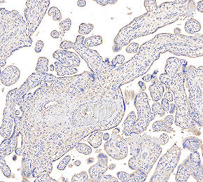 Immunohistochemical analysis of formalin fixed paraffin embedded human placenta tissue with F2400 at 1:50 dilution.
Usage Information
| Dilution |
|---|
|
| Application |
|---|
| IHC, FCM |
| Reactivity |
|---|
| Human |
| Source |
|---|
| Mouse Monoclonal Antibody |
| Storage Buffer |
|---|
| PBS, pH 7.2+50% Glycerol+0.05% BSA+0.01% NaN3 |
| Storage (from the date of receipt) |
|---|
| -20°C (avoid freeze-thaw cycles), 2 years |
| Positive Control | Human placental; HepG2 |
|---|---|
| Negative Control |
Experimental Methods
| IHC |
|---|
Experimental Protocol:
Deparaffinization/Rehydration
1. Deparaffinize/hydrate sections:
2. Incubate sections in three washes of xylene for 5 min each.
3. Incubate sections in two washes of 100% ethanol for 10 min each.
4. Incubate sections in two washes of 95% ethanol for 10 min each.
5. Wash sections two times in dH2O for 5 min each.
6.Antigen retrieval: For Citrate: Heat slides in a microwave submersed in 1X citrate unmasking solution until boiling is initiated; continue with 10 min at a sub-boiling temperature (95°-98°C). Cool slides on bench top for 30 min.
Staining
1. Wash sections in dH2O three times for 5 min each.
2. Incubate sections in 3% hydrogen peroxide for 10 min.
3. Wash sections in dH2O two times for 5 min each.
4. Wash sections in wash buffer for 5 min.
5. Block each section with 100–400 µl of blocking solution for 1 hr at room temperature.
6. Remove blocking solution and add 100–400 µl primary antibody diluent in to each section. Incubate overnight at 4°C.
7. Remove antibody solution and wash sections with wash buffer three times for 5 min each.
8. Cover section with 1–3 drops HRPas needed. Incubate in a humidified chamber for 30 min at room temperature.
9. Wash sections three times with wash buffer for 5 min each.
10. Add DAB Chromogen Concentrate to DAB Diluent and mix well before use.
11. Apply 100–400 µl DAB to each section and monitor closely. 1–10 min generally provides an acceptable staining intensity.
12. Immerse slides in dH2O.
13. If desired, counterstain sections with hematoxylin.
14. Wash sections in dH2O two times for 5 min each.
15. Dehydrate sections: Incubate sections in 95% ethanol two times for 10 sec each; Repeat in 100% ethanol, incubating sections two times for 10 sec each; Repeat in xylene, incubating sections two times for 10 sec each.
16. Mount sections with coverslips and mounting medium.
|
Biological Description
| Specificity |
|---|
TGF β Receptor II Antibody [J15L22] detects endogenous levels of total TGF-β Receptor II protein. |
| Subcellular Location |
|---|
| Cell membrane, Membrane, Secreted |
| Uniprot ID |
|---|
| P37173 |
| Clone |
|---|
| J15L22 |
| Synonym |
|---|
| TGF-beta receptor type-2, TGFR-2, TGF-beta type II receptor, Transforming growth factor-beta receptor type II, TGF-beta receptor type II, TbetaR-II, TGFBR2 |
| Background |
|---|
| The TGF β Receptor II, (transforming growth factor-β type II receptor, TβRII) is a type I transmembrane glycoprotein and a constitutively active serine/threonine kinase that initiates TGF-β signaling. It comprises an extracellular ligand-binding ectodomain with a three-finger toxin fold stabilized by six disulfide bonds (four conserved among type II receptors and two unique to TβRII), a single transmembrane segment, and an intracellular kinase domain. The ectodomain contains 12 cysteines and features an extended first “finger” compared to related receptors, enabling specific ligand interactions. TβRII is expressed in many tissues and binds TGF-β ligands, recruiting and phosphorylating the type I receptor (TβRI) to activate SMAD-dependent and SMAD-independent pathways, including Src kinase signaling. Through these mechanisms, TβRII regulates development, tissue homeostasis, immune responses, and pathological processes such as cancer and fibrosis. |
| References |
|---|
|
Tech Support
Tel: +1-832-582-8158 Ext:3
If you have any other enquiries, please leave a message.
Products are for research use only. Not for human use. We do not sell to patients.
©Copyright 2013 Selleck Chemicals. All Rights Reserved.