research use only
2-Methoxyestradiol (2-MeOE2) HIF inhibitor
Cat.No.S1233
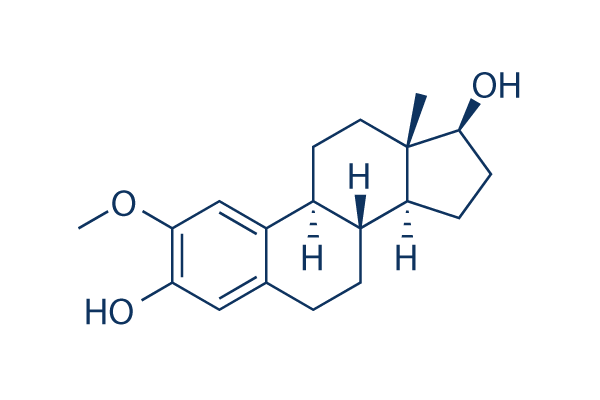
Chemical Structure
Molecular Weight: 302.41
Quality Control
| Related Targets | EGFR VEGFR JAK PDGFR FGFR Src FLT FLT3 HER2 Bcr-Abl |
|---|---|
| Other HIF Inhibitors | PT2399 PX-478 Dihydrochloride BAY 87-2243 KC7F2 Lificiguat (YC-1) IOX2 CAY10585 (LW 6) Molidustat (BAY 85-3934) PT2385 IDF-11774 |
Cell Culture, Treatment & Working Concentration
| Cell Lines | Assay Type | Concentration | Incubation Time | Formulation | Activity Description | PMID |
|---|---|---|---|---|---|---|
| HOP-62 | cytotoxicity assay | ~100 μM | GI50=0.70 μM | |||
| HCT-116 | cytotoxicity assay | ~100 μM | GI50=0.47 μM | |||
| SF-539 | cytotoxicity assay | ~100 μM | GI50=0.32 μM | |||
| UACC-62 | cytotoxicity assay | ~100 μM | GI50=0.36 μM | |||
| OVCAR-3 | cytotoxicity assay | ~100 μM | GI50=0.21 μM | |||
| SN12C | cytotoxicity assay | ~100 μM | GI50=0.95 μM | |||
| DU-145 | cytotoxicity assay | ~100 μM | GI50=1.8 μM | |||
| MDA-MB-435 | cytotoxicity assay | ~100 μM | GI50=0.08 μM | |||
| LNCaP | Growth inhibitory assay | ~50 μM | IC50=0.5 μM | |||
| DU145 | Growth inhibitory assay | GI50=1.22 μM | ||||
| MDA-MB-23 | Growth inhibitory assay | GI50=0.94 μM | ||||
| MCF7 | Growth inhibitory assay | GI50=2.35 μM | ||||
| U87-MG | Growth inhibitory assay | IC50=8.54 μM | ||||
| PC3 | Growth inhibitory assay | IC50=2.65 μM | ||||
| HUVEC | Growth inhibitory assay | IC50=0.84 μM | ||||
| U937 | Growth inhibitory assay | IC50=2.91 μM | ||||
| SK-OV-3 | Function assay | circumvents Pgp-mediated drug resistance with EC50 of 867 nM | ||||
| SK-OV-3 MDR-1-6/6 | Function assay | circumvents Pgp-mediated drug resistance with EC50 of 2268 nM | ||||
| HeLa | Function assay | inhibitsβIII-tubulin on drug sensitivity | ||||
| HUVEC | Function assay | shows antiangiogenic activity | ||||
| Click to View More Cell Line Experimental Data | ||||||
Chemical Information, Storage & Stability
| Molecular Weight | 302.41 | Formula | C19H26O3 |
Storage (From the date of receipt) | |
|---|---|---|---|---|---|
| CAS No. | 362-07-2 | Download SDF | Storage of Stock Solutions |
|
|
| Synonyms | NSC 659853, 2-ME2 | Smiles | CC12CCC3C(C1CCC2O)CCC4=CC(=C(C=C34)OC)O | ||
Solubility
|
In vitro |
DMSO
: 60 mg/mL
(198.4 mM)
Water : Insoluble Ethanol : Insoluble |
Molarity Calculator
|
In vivo |
|||||
In vivo Formulation Calculator (Clear solution)
Step 1: Enter information below (Recommended: An additional animal making an allowance for loss during the experiment)
Step 2: Enter the in vivo formulation (This is only the calculator, not formulation. Please contact us first if there is no in vivo formulation at the solubility Section.)
Calculation results:
Working concentration: mg/ml;
Method for preparing DMSO master liquid: mg drug pre-dissolved in μL DMSO ( Master liquid concentration mg/mL, Please contact us first if the concentration exceeds the DMSO solubility of the batch of drug. )
Method for preparing in vivo formulation: Take μL DMSO master liquid, next addμL PEG300, mix and clarify, next addμL Tween 80, mix and clarify, next add μL ddH2O, mix and clarify.
Method for preparing in vivo formulation: Take μL DMSO master liquid, next add μL Corn oil, mix and clarify.
Note: 1. Please make sure the liquid is clear before adding the next solvent.
2. Be sure to add the solvent(s) in order. You must ensure that the solution obtained, in the previous addition, is a clear solution before proceeding to add the next solvent. Physical methods such as vortex, ultrasound or hot water bath can be used to aid dissolving.
Mechanism of Action
| Targets/IC50/Ki |
HIF-2α
(Rat aortic smooth muscle A-10 cells) Microtubule Associated
(Cell-free assay) HIF-1α
(MDA-MB-231 cells) |
|---|---|
| In vitro |
2-Methoxyestradiol (2-MeOE2) exhibits the inhibitory activity of cellular proliferation in a breast carcinoma cell line MDA-MB-435 and an ovarian carcinoma cell line SK-OV-3 with IC50 of 1.38 μM and 1.79 μM, respectively. Furthermore, it also inhibits cellular microtubule depolymerization in rat aortic smooth muscle A-10 cells with EC50 of 7.5 μM. This compound inhibits proliferation of MCF-7 and BM cells with IC50 of 52 μM and 8 μM. In MDA-MB-231 cells, it inhibits HIF-1-mediated transcriptional activation of target genes without affecting the transcription of HIF-1α itself. A recent study shows that 2-MeOE2 (0.5 μM), blocks TGF-β3-induced expression of collagen (Col) type I(αI), Col III(αI), plasminogen activator inhibitor (PAI) 1, connective tissue growth factor (CTGF), and α-smooth muscle actin (α-SMA). Moreover, it ameliorates TGF-β3-induced Smad2/3 phosphorylation and nuclear translocation, and inhibits TGF-β3-induced activation of the PI3K/Akt/mTOR pathway. |
| Kinase Assay |
Microtubule depolymerizing activity
|
|
The effects of 2-Methoxyestradiol (2-MeOE2) on cellular microtubule depolymerization are determined by indirect immunofluorescence techniques in rat aortic smooth muscle A-10 cells. Microtubules are visualised using a β-tubulin antibody. Three viewers determine the percent microtubule loss for each treatment concentration. The data are averaged and plotted as percent microtubule loss versus drug concentration, and the EC50s for microtubule depolymerization are calculated from the log dose–response curves.
|
|
| In vivo |
In a 9L rat glioma (9L-V6R) rat model, 2-Methoxyestradiol (2-MeOE2) significantly decreases HIF-1 activity and inhibits tumour growth in a dose-dependent manner, with a 4-fold reduction at 60 mg/kg/day and a 23-fold reduction at 600 mg/kg/day, respectively. |
References |
|
Tech Support
Tel: +1-832-582-8158 Ext:3
If you have any other enquiries, please leave a message.
Products are for research use only. Not for human use. We do not sell to patients.
©Copyright 2013 Selleck Chemicals. All Rights Reserved.